Imalytics Preclinical
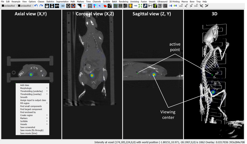
Imalytics Preclinical is a software for interactive segmentation, reconstruction, 3D-visualization, and analysis of biological and medical image data sets using interactive segmentation operations.
The software is preferably used for fast and accurate analysis of single-modality images such as preclinical μCT or clinical CT data as well as for the analysis of multimodal image data such as PET-CT, FMT-CT, SPECT-CT, MRI-CT, PET-SPECT, etc.
Furthermore, it is suitable for the analysis of ultrasound (US), bioluminescence, and magnetic resonance imaging (MRI) data. Depending on the imaging modality, 2D, 3D, and 4D data (dynamic images) can be processed.
Analysis of cells, tissues, organs and whole-body scans for insects, fish, birds, mice, rats, sheep, other mammals, and even humans can be performed, but the software is not approved for clinical use yet.
The software supports many different applications and undergoes continuous improvement and optimization processes. It has successfully been used for over 80 scientific publications at different research institutes, such as Aachen, Roche, Harvard, Jena, Düsseldorf, and Frankfurt.
Details of the software are described in this publication:
F. Gremse, et al., “Imalytics Preclinical: Interactive Analysis of Biomedical Volume Data,” Theranostics, vol. 6, no. 3, pp. 328–341, 2016.
A video showing the software has ben published here:
F. Gremse et al., “Hybrid μCT-FMT imaging and image analysis,” J Vis Exp, no. 100, p. e52770, Jun. 2015.