Applications
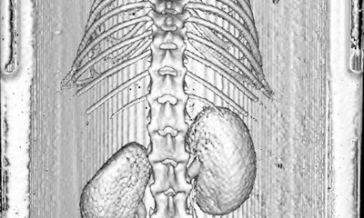
Image Processing
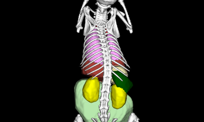
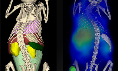
Imalytics Preclinical allows processing and visualization of (i) an underlay (e.g. a CT image), a segmentation map (e.g. whole body organ segmentation), and an overlay (e.g. three-dimensional fluorescence distribution).
3D Printing
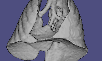
With Imalytics Preclinical different isosurfaces such as organs, the skeleton, the whole mouse body, or the mouse bed of an image can be exported as a stl-file for 3D printing.
Spectral Unmixing
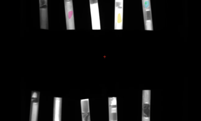
To analyze multispectral images, it is possible to perform a spectral unmixing using Imaltics Preclinical.
Segmentation
Segmentation of organs or regions of interest (ROI), even whole body organ segmentation are easy to conduct with Imalytics Preclinical.
Quantification
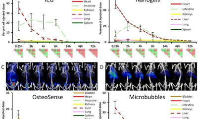
Signal intensities in ROIs, organs, or the whole object can be easily quantified using the batch function of Imalytics Preclinical. Thereby, several objects and points in time can be analysed in one single step.
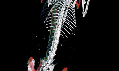
Image Fusion
Images from different modalities can be fused using manual or marker-based alignment in Imalytics Preclinical making multimodal image data analysis straightforward.
Multimodal Imaging
One of our main key expertise lies in fusion, processing, and analysing multimodal image data such as optical-CT, nuclear-CT, MRI-optical, or MRI-CT data sets.
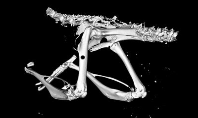
Bone Analysis
The analysis of different bone structures and features, the evaluation and quantification of new bone formation as well as the occurrence of calcification were integrated into the software.
More Information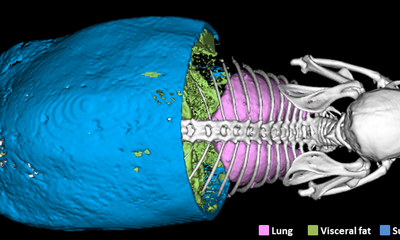
Fat analysis
Imalytics Preclinical is intensively used for determining whole body fat, differentitaion into subcutaneous and visceral fat, and segmentation of brown adipose tissue.
More Information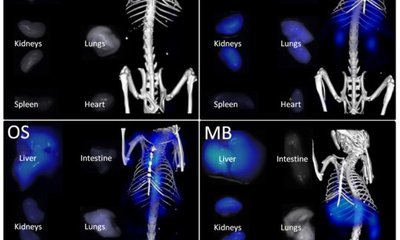
Pharmacokinetic
Our software and developed kinetic models has been widely used to determine the organ biodistribution, elimination, and retention sites of newly developed nanocarriers or drug delivery systems.
More Information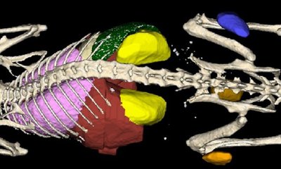
Cancer research
Imalytics Preclinical offers the possibility to accurately detect, segment, and characterize tumors, lesions, and metastases. It was already used for colon cancer, PDAC, liver tumors and lesions, lung metastases, and several types of subcutaneous and orthotopic tumors.
More Information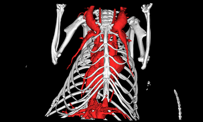
Vascular analysis
The software has been successfully used to assess the vascular system and the changes in the blood vessels such as atherosclerotic inflammation, lesions or calcifications, and stenosis in the carotids.
More Information