Cancer Research
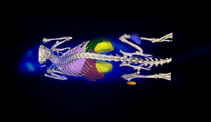
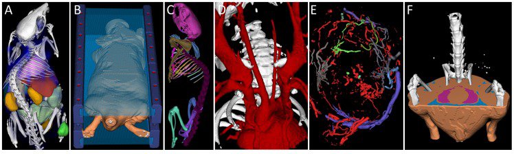
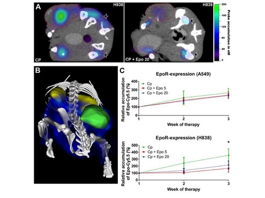
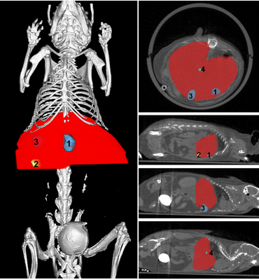
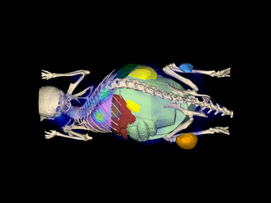
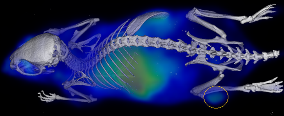
Our analysis software was intensively used for the detection, segmentation of tumors and metastases. Their exact localization and extension can be visualized in 3D, and also their volume and diameter can be calculated. Depending on the use of a contrast agent, information about blood flow, vascularization, and neo-angiogenesis (after treatment) can be obtained within the tumors and metastases.
On the picture gallery above you find representative images of subcutaneous tumors derived from different cell types, liver metastasis (1-4), and segmented tumor blood vessels (E).